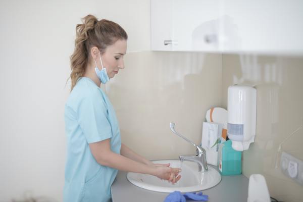
Blog
Haglund’s Deformity (also called Bauer bump or pump bump)
This type of deformity looks like an enlargement of the bone (the bone is called calcaneus) just above and behind the heel. There is no swelling at the site, only pain and/or soreness. If a person wears shoes with a tight heel counter (the part of the shoe around the heel), then they aggravate it so wearing sandals is more comfortable.
Haglund’s deformity is caused by an inflexible heel counter that is often present in ice skates and high-heeled shoes. This is when the bony prominence (exostosis) begins to develop; soreness develops only later.
This type of deformity can lead to retrocalcaneal bursitis (inflammation of the bursa behind the calcaneus). Furthermore, the prominence itself leads to more bursitis by irritating it. In fact, Haglund’s deformity is often misdiagnosed as retrocalcaneal bursitis. While both conditions have the symptoms of irritation, redness, and enlargement at or near the base of Achilles tendon, Haglund’s deformity is painful only when pressed and has redness and swelling that does not spread further than the prominence itself.
X-ray images help to diagnose Haglund’s deformity and MRI can show its outline.
To help with the pain upon pressure and reduce the strain on Achilles tendon, one can wear shoes without heels or with stretched, cushioned or softer heel counters. Also, the heels of the shoes can be modified with a 6 mm heel-raise, which can be done by a certified pedorthist.
The common treatment is PRICEMM: Protection, relative Rest, Ice, Compression, Elevation, Medication, and rehabilitation exercise Modalities. It’s also useful to complete an eccentric strengthening program and perform soleus and gastrocnemius stretching. If this treatment has not helped, after 6 months surgery may be recommended.
Ankle Ligament Sprains
Ankle ligaments hold the joint and bones together in a joint called a talocrural joint. The bones that are held together here are lower tibia and fibula of the lower legs, as well as talus, a small bone that fits between them. This joint is like a hinge: it moves allowing the toes to lift off the ground (dorsiflexion) and swings down to plant the foot firmly on the ground (plantarflexion). The list of ligaments of the foot that are commonly injured can be found in the previous blog entry. If the foot is rotated outward during a sprain, it’s called an eversion sprain. It comprises 5-10% of all ankle sprains and takes much longer to heal than an inversion sprain. An inversion sprain is much more common.
Ankle sprains are classified into Grades 1, 2, & 3, depending on their severity. Grade 1 is a mild sprain. There is some pain and very little swelling because the ligament has been stretched but there is no deformity; return to normal function can be expected in 2 weeks. Grade 2 is when the ligament is partially torn. Some bleeding and bruising can occur and is often lower than the injury site due to gravity. Here rehabilitation is necessary and the person can return to normal activities within 2-6 weeks. Grade 3 sprain is a complete tear of a ligament. Pain is severe at first but then subsides quicker than a Grade 2 sprain. Surgery could be necessary and return to activity can take as long as 6 weeks or more.
Common testing includes Ottawa ankles rules: if pain is present near the malleoli (the two bones protruding on the inside and outside of the ankle) and the person cannot walk for 4 steps after injury, an X-ray should be ordered. Treatment includes avoiding HARM (heat, alcoholic beverages, running, and massage). Ultrasound can be indicated to reduce swelling only if the person is 18 years old or older. Casting a sprained ankle is not recommended. Use the acronym PRICE (protection, rest, ice, compression, and elevation).
There are many different types of ankle braces that can provide protection. An ankle can be taped to provide support short-term. If there is a severe sprain, a short-leg cast or a walking boot can be used to partially immobilize the leg but not completely. Elevate the ankle and apply ice-packs wrapped in a wet towel for 15-20 minutes every 2 hours for the first 2-3 days. Compressive wrap should be applied correctly to ensure adequate blood flow to the toes. Once the swelling begins to subside, your MD will refer you to a physiotherapist whose advice the client should follow as precisely as possible. Gradually begin to move the ankle 48 hours after the sprain as part of rehabilitation. Make sure footwear is roomy enough to accommodate swelling and it gives enough ankle support through a wide stable base. The soles of the shoes should not be worn down especially at the heel causing ankle instability.
Chronic Ankle Instability (CAI)
Clients describe CAI as a feeling of “giving way” and instability resulting in swelling, prolonged pain, increased or (if ankle sprain has previously occurred) limited range of motion. CAI often leads to ankle injuries and, most commonly, ankle sprains. The three most common ankle sprains are: inversion sprains (the sole of the foot is rotated inward to face the other foot), eversion sprains (the rotation is outward and usually affects deltoid ligaments), and high ankle sprains (harm to the syndesmotic ligaments). 75% of all ankle injuries are sprains and 85% of all ankle sprains are inversion sprains, which tend to recur. People most affected by CAI are usually physically active. Those that tend to have lax ankle ligaments (mechanical issue) and have an active lifestyle (functional issue) tend to suffer from CAI the most. Those with a cavus foot (high arch of the sole of the foot) are also more likely to have CAI, particularly, lateral ankle instability. If the joint is stable, intact and ligaments are strong, injury is less likely to occur.
Anterior talofibular ligament is the most commonly injured and also the weakest ankle ligament. ATFL inversion sprain can occur when a foot is inverted and plantarflexed (the position of the foot when toes are pointed out) while the leg is externally rotated (leg is turned to point to the side). The next most common sprain is a tear in the calcaneofibular ligament (CFL) often followed by a tear of the AFTL. A sprain of the posterior talofibular ligament (PTFL) of the outer side of the ankle is least common of the three because it’s the third weakest ligament. The medial (inner or facing the midline) ligaments are: tibiocalaneal, tibionavicular, superficial tibiotalar, and posterior tibiotalar ligaments and are least likely to be sprained.
As part of the assessment, the physician would often order stress radiographs and X-rays. To estimate loss or return of functional ankle control and predict future ankle injury postural control is used. Postural control is one’s ability to keep centre of mass centred over one single foot. Most commonly, to improve postural control taping, bracing, and orthotics are used. In extreme cases, the physician may recommend reconstructive surgery of the ankle.
Meniscal Injuries, etc… What is a Meniscus, anyway?
Meniscus, also called semi-lunar cartilage (rubber-like padding around bones where they join together), is knee-joint cartilage which grows inward from the joint capsule and forms pads or discs. These discs are situated within the knee to cushion the contact between femur (the bone of upper leg) and tibia (the biggest bone of the lower leg). They deepen tibia’s surface and provide a larger and more stable surface area for the femur to join with. Their function is shock absorption, reducing friction, and stabilization of the knee joint.
Half of all knee ligament and meniscal injuries are sports injuries. There are chronic and acute meniscal tears. Chronic degenerative tears rarely cause any dysfunction or pain since they are caused by many episodes of minimal trauma. When constant shearing forces act on the inner meniscus, it may lead to detachment, pain, instability, and momentary locking. Horizontal cleavage tears are chronic in nature and are due to degenerative changes in the back inner part of the knee.
The three acute tears are: longitudinal, bucket-handle, and parrot-beak, and are classified by their shape. The most common are longitudinal tears and happen when the knee is twisted while flexed and the foot is fixed in one place. Bucket-handle tears have 40% frequency of incidence. They happen when an entire longitudinal segment of meniscus is displaced toward the center of the tibia, often leading to the knee locking in place when flexed. Parrot-beak tears are often found in adolescents and involve the mid-outer meniscus developing two tears in a parrot-beak shape.
Immediate treatment includes RICE (rest, ice, compression, elevation), non-steroidal anti-inflammatory medications (NSAIDs), and crutches to keep the weight off the injured knee. See your family health professional immediately if the knee is locked in place or if excessive joint effusion (abnormal accumulation of fluid in a joint) is present. Physiotherapy, surgery, and/or orthoses (braces or other devices that support and correct joint alignment) may be prescribed by family doctor or nurse practitioner.
Quality Foot Care for Those with Morton’s Neuroma
If you are a fashion-minded woman ages 30 to 60, you are at a high risk of developing Morton’s Neuroma. This is a type of nerve pain that often occurs in the third intermetatarsal space (in the space where the 3rd and 4th toes join together) but can occur in other intermetatarsal spaces also. The frequency of Morton’s Neuroma is directly related to how one walks and by the anatomical structure of the foot. The pain is caused by a lesion or injury to one of the common digital (toe) nerves due to repeated trauma or compression, often when wearing high-heeled and narrow shoes. That’s why, this condition is more common in women. The pain is often worse when walking but can occur at rest. Another symptom is loss of sensation in the involved toes and affected toes may be splayed.
The most effective initial treatment is, commonly, a Neuroma Pad placed in shoes with large enough toe-box. Make sure to wear footwear with deep and wide toe-box, low heels, and stiff soles. If a neuroma pad does not provide enough pain relief, use of professional orthotics is recommended. In extreme cases, Morton’s Neuroma can be treated with ultrasound or laser treatment.
Osteoarthritis
It’s a degenerative disease of joints that involves cartilage, bone, and synovia. With wear and friction, cartilage that surrounds the bones at a joint thins, becomes rough, and causes pain, deformity, and instability. Unstable joints often buckle or lock in place, especially the knee. Tenderness, stiffness, and pain of the joint, not getting relief from over-the-counter medications, pain changes with weather, joint swelling, restricted movement, creaking and grating, and bony enlargement are common symptoms. Pain in the joint develops gradually, worsens with use and is relieved with rest. Pain and stiffness increase with damp weather and low air pressure because nerve fibres in the capsule of the knee are sensitive to changes in atmospheric pressure. An X-ray is used to diagnose osteoarthritis. It affects more women than men. Mild to moderate pain can be treated with non-steroidal anti-inflammatory drugs, such as aspirin or ibuprofen. Other treatments may include surgical and non-surgical therapies, such as orthotics, well-cushioned shoes, and physiotherapy. A family doctor would make the diagnosis and may refer the client to a podiatrist or chiropodist.
Women’s Plantar Fasciitis Orthotic Insole (at Mark’s Work Warehouse)
Did you know? If you have Plantar Fasciitis, inflammation of the plantar fascia, you can: use RICE (rest, ice, compression, elevation) and tape the afflicted area for 1 to 5 days to achieve pain relief and support. Then, wean into orthotics and/or shoes with good arch support. Also, support pes planus or pes cavus and use anti-inflammatory medications for pain. The use of TENS and ultrasound are also recommended.
Diabetic Foot Ulcers
This type of ulcer is a complication of Diabetes. It’s a break in skin integrity where harmful bacteria can easily invade and multiply. The cause is often peripheral neuropathy or peripheral vascular disease. The ulcers most commonly associated with Diabetes are neuropathic, arterial and venous.
Neuropathic ulcers are caused by frequent friction on parts of the foot where there is the most weight. It often occurs on the bottom of a great toe or 1st metatarsal head. This type of ulcer is often painless, surrounded by a callous, and round in shape.
Ischemia, or decreased arterial blood flow to the feet causes arterial ulcers. These are most serious and are often located on the heels, tips of toes, between toes, sides or soles of feet, lateral malleoli, and metatarsal heads.
Venous ulcers happen due to lack of return of venous blood flow to the heart and accumulation of this blood in the lower legs. This makes the skin dry, itchy, dark in colour, swollen, and flakey. The site of venous ulcers is often on the lower legs and inside of the ankles.
Footwear that does not fit is a big cause of foot ulcers. Things to watch out for to prevent ulcers are: decreased circulation and lack of sensation.
New Research on Treatment of Plantar Warts
Over the counter preparations that include Salycylic acid, dichloroacetic or trichloroacetic acids are most affective. But never put this preparation on intact skin. Use a piece of adhesive tape (ex. Leukoplast) and cut a hole in the middle for the wart to isolate it. Apply the preparation to the wart and cover with another piece of tape. Keep it there dry for one week. The wart can then be shaved down. The whole process can be done again on a weekly basis until the wart is gone. Cryotherapy, which is freezing the wart with liquid nitrogen, is also effective but must be done by the physician. If you have diabetes or peripheral vascular disease Do Not shave down (pare) the wart.
Reference
emedicinehealth.com